Further Contributions to the Solution
of the Piltdown Problem ss
J. S. Weiner; W. E. Le Gros Clark; K. P. Oakley; G. F. Claringbull & M. H. Hey; F. H. Edmunds; S. H. U. Bowie & C. F. Davidson; C. F. M. Fryd; A. D. Baynes-Cope; A. E. A. Werner & R. J. Plesters
Bulletin of the British Museum (Natural History) Geology 1955
CONTENTS
Page
Introduction. Sir Gavin de Beer, F. R. S. 228
I. Outline of the Piltdown Problem. J. S. Weiner 229
2. An Anatomical Study of the Piltdown Teeth and the so-called Turbinal Bone.
W. E. Le Gros Clark, F.R.S. 234
3. The Piltdown "Implements." K. P. Oakley 243
4. The Piltdown Mammalia. K. P. Oakley 247
5. The Composition of the Piltdown Hominoid Remains. K. P. Oakley 254
6. Chemical Changes in Bones: a Note on the Analyses. C. F. M. Fryd 266
7. The X-ray, Crystallography of the Piltdown Fossils. G.F.Claringbull &
M. H. Hey 268
8. The Black Coating on the Piltdown Canine. A. E. A. Werner & R. J. Plesters 271
9. The Geology of the Piltdown Neighbourhood. F. H. Edmunds 273
10. The Radioactivity of the Piltdown Fossils. S. H. U. Bowie & C. F. Davidson 276
11. The Fluorimetric Determination of Uranium in the Piltdown Fossils.
A. D. Baynes-Cope. 283
12. References 185
TABLES:
I. Composition of Stains on Piltdown Flints 245
II. Radioactivity of Piltdown Elephant teeth and other Villafranchian Fossils 249
III-IV. Analyses of Piltdown Hominoid Bones and Teeth 262
V. . Analyses of Piltdown Mammalian Bones and Teeth 263
VI-VII. Analyses of Bones and Teeth used in comparison 264-265
VIII-IX. X-ray Examination of Piltdown Specimens and of Bones Artificially
Iron-stained 269-270
X-Xl. Radiometric Assays of Piltdown Specimens and various Tertiary,
Pleistocene and Holocene fossils 279-282
XII. Uranium Content of Piltdown and other Fossils 284
PLATES:
27. Piltdown Mandible and Orang Mandible compared
28. Piltdown Flint "Implements"
29. Piltdown Bone "Implement"
30. Electron-micrograph of Piltdown Mandible and Autoradiograph of Piltdown
Elephant Molar
31. X-ray Diffraction Photographs of Apatite and Gypsum in Piltdown and other Bones
INTRODUCTION
[228] We are now in a position to give an account of the full extent of the Piltdown hoax. The mandible has been shown by further anatomical and X-ray evidence to be almost certainly that of an immature orang-utan; that it is entirely Recent has been confirmed by a number of microchemical tests, as well as by the electron-microscope demonstration of organic (collagen) fibres; the black coating on the canine tooth, originally assumed to be an iron encrustation, is a paint (probably Vandyke brown); the so-called turbinal bone is shown by its texture not to be a turbinal bone at all, but thin fragments of probably non-human limb-bone; all the associated flint implements have been artificially iron-stained; the bone implement was shaped by a steel knife; the whole of the associated fauna must have been "planted," and it is concluded from radioactivity tests and fluorine analysis that some of the specimens are of foreign origin. The human skull fragments and some of the fossil animal bones are partly replaced by gypsum, the result of their treatment with iron sulphate to produce a colour matching that of the gravel. Not one of the Piltdown finds genuinely came from Piltdown. These latest investigations have demonstrated the methods now available which will not only make a successful repetition of a similar type of forgery virtually impossible in the future, but will be of further value in palaeontological research.
Gavin de Beer,
Director.
[234] 2. An Amatomical Study of the Piltdown Teeth and the So-called Turbinal Bone
By W. E. Le Gros Clark, F.R.S.
Department of Anatomy, University of Oxford
(I) The Anatomical Details of the Piltdown Teeth
The teeth from Piltdown are the first and second right lower molars in the mandibular fragment an isolated canine tooth, and an isolated left lower molar tooth. The last was reported to have been found in a heap of stones raked off a ploughed field about two miles from the original Piltdown site. Considered by themselves, it seems certain that the teeth would have been attributed to an anthropoid ape (quite similar to a chimpanzee or an orang) except for two main features : (i) the extremely flat wear of the molar teeth in the mandible, which is not normally to be found in pongid teeth at an equivalent stage of attrition, but which closely approximates to the type of wear commonly found in hominid molars; and (2) the quite unusual type of wear on the canine which, so far as we are aware, is not paralleled in the canines of any of the known genera of anthropoid apes, recent or extinct. The suggestion that these aberrant features might be the result of artificial abrasion at once offered a plausible explanation of their seemingly anomalous character. Indeed, it may well be asked why such a suggestion had not been seriously considered until quite recently. There are no doubt several reasons for this. In the first place, the mandibular fragment and the canine tooth were reported to have been found by experienced palaeontologists during their excavations at Piltdown, and the occurrence in situ has thus always been accepted without question. Secondly, the faking, obvious though it now appears, had been accomplished with extraordinary skill : and, lastly, the statement that the worn surface of the canine shows an exposure of secondary dentine would almost certainly have distracted attention from a possible consideration of faking by artificial abrasion. Secondary dentine is deposited as a reaction to prolonged natural wear and its presence in the canine would thus presuppose that the excessive wear of this tooth was indeed natural. In fact, a reexamination of the canine has shown that there is no evidence of the deposition of secondary dentine.
Two other relevant features have been held by some authorities to distinguish the Piltdown molars of the mandible from those of anthropoid apes. One is their hypsodont character. But comparative study has shown that, while a similar degree
[235]
Text-Figure 2 Text- Figure 4
of hypsodonty is very unusual (if, indeed, it does occur) in chimpanzees and gorillas, it is not uncommon in the orang. The other feature is the relative shortness of the roots of the molar teeth as they appeared to be displayed in the original radiograph of the Piltdown molars published in a paper by Underwood (1913) and subsequently copied in publications by other authors (e.g. Keith, 1915; Lyne, 1916). But new radiographs taken recently show quite clearly that the roots are in fact markedly longer than they have been portrayed, and are thus entirely simian in appearance. This seems to us to be an important point which needs to be emphasized. The original radiographs lacked sufficient definition to outline the roots distinctly; from the recently taken radiograph the outlines of the molars have been reproduced for comparison with those made from appearances shown in the original radiograph (Text-figs. 1, 2). It will be observed that the lower end of the anterior root of M1
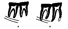
Text-Fig.. I. A. Outline drawing of the molar teeth in the Piltdown mandible showing the roots as they had been interpreted on the basis of the original radiograph published in 1913. B. A similar drawing made from a recent radiograph showing the actual form and extent of the roots. The anterior root of M1 has been broken off, and its probable extent is indicated by a broken line. In both figures the position of the mandibular canal is shown. Natural size.

Text-Fig. 2. Radiograph of the two molar teeth in the Piltdown mandible showing the form and extent of their roots. Note the apparently accurate apposition of the crowns of the teeth at their contact facets. Twice natural size. [X-ray by P. E. Purves.]
[236] has been broken off (presumably it has been involved in the fracture of the mandible at this level). Its total length can therefore not be precisely estimated, but there is an indication of the lower end of the socket which suggests that originally the apex of the root curved downwards and backwards for some distance. The posterior root of M1 is long and is defected backwards at its lower extremity. The radiograph of the mandible shows that it actually reaches the upper border of the mandibular canal. The anterior root of M2 is well defined and was correctly displayed in the original radiograph. Compared with the posterior root of M1, the rounded and blunt apex of this root and the relative width of the apical canal suggest that the root had perhaps not completed its full development. The posterior root of M2 is considerably longer than the anterior root, extending downwards to the level of the lower border of the mandibular canal. This relationship is of some significance, for the "hominid" appearance in the original radiograph showing the roots of both the first and second molars apparently falling well short of the canal certainly misled some authorities. Like the anterior root, the apex of the posterior root of M2 is bluntly rounded and the apical canal relatively wide.
The problem having been posed–is the unusual type of wear of the Piltdown molar teeth the result of natural attrition during life or of artificial abrasion after death?–consideration was given to those details which on close inspection might be expected to differentiate the one from the other. A critical study of the teeth at once revealed certain features which had either escaped notice previously, or the possible significance of which had not been realized. Indeed, it was because these features appeared to lend such strong support to the hypothesis of artificial abrasion that it was decided to re-examine all the Piltdown material for further evidence of faking.
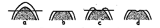
Text-Fig. 3. Diagram illustrating the peculiar type of abrasion on the cusps of the Piltdown molar teeth. In (a) is shown a schematic section through an unworn cusp. If this were subjected to artificial abrasion in the plane indicated, the appearance shown in (b) would be produced, i. e., a flat area of exposed dentine (stippled) flush with the surrounding enamel; such an appearance is seen on the antero-internal cusps of the Piltdown molars. In (c) is shown a schematic section through a cusp partially worn by natural attrition, illustrating the concavity of the dentine depressed below the surrounding enamel. Artificial abrasion in the plane indicated would produce the appearance shown in (d), i.e. a depressed "dimple" in the centre of a flat area of dentine flush with the surrounding enamel; such an appearance is seen on the antero-external cusps of the Piltdown molars.
The flatness of the molar teeth is astonishingly even over almost the entire extent of the occlusal surface, as though, indeed, the latter had been planed down by some rapidly acting shearing force. A considerable area of dentine (about 4 mm. in its greatest diameter) has been exposed on the antero-internal cusps of both teeth, and not only are these areas quite flat, they are also flush with the surrounding enamel (Text-fig. 3, a, b). But in natural attrition, whether in hominids or pongids, areas [237] of dentine exposed to this degree from shallow concavities, because the less hard dentine wears away more rapidly than the surrounding and harder enamel. The possibility that the appearance of the occlusal surfaces of the Piltdown molar teeth might be the result of an unusual type of natural attrition, which had proceeded so rapidly that there was insufficient time for the exposed dentine to be hollowed out below the level of the surrounding enamel, does not seem an acceptable explanation. In the first place we have not been able to find, in an examination of 137 ape jaws and 200 human jaws, a parallel condition in areas of dentine of similar extent exposed by natural attrition. Secondly, the small punctate exposures of dentine on the external cusps of the Piltdown molars are in fact already depressed below the surrounding enamel, presumably as the result of natural wear (and thus indicating that the latter was originally of the normal type). Moreover, on the antero-external cusp of both molars the punctate exposure of dentine is surrounded by a small and perfectly flat area of dentine which, it appears, must have been exposed (like the areas of dentine on the antero-internal cusps) by artificial abrasion (Text—fig.3, c, d). These flat areas of dentine are stained a dark brown colour and are sharply defined by a thin outline of deeper staining. We have been able to produce outlines of somewhat similar appearance in modern teeth which have been filed down and artificially iron stained, and it seems probable that the outline of deeper staining may be due to the relatively rich amount of organic substance at the dentino-enamel junction, where there is a greater proportion of interprismatic substance to enamel-rods (Noyes, 1948), and also to the considerable degree of branching and interlacing of dentinal tubules at the surface of the dentine.
Another curious feature of the molars of the Piltdown mandible is that the dentine on both M1 and M2 is much more extensively exposed on the antero-internal than the antero-external cusps. But this is the reverse of natural wear, in which the outer cusps of the lower molars are normally worn down more rapidly in the earlier stages of attrition. Out of 200 human jaws there were only 4 in which (at an approximately equivalent stage of wear) the dentine of a lower molar was exposed to an approximately equal extent on the outer and inner cusps; in one other the area of exposed dentine on the inner cusp was rather more extensive (though by no means to the degree shown in the Piltdown teeth). Thus with normal occlusion it must be at least very exceptional to find human teeth in which the wear on the antero-internal cusps is so much greater than on the antero-external cusps of M1 and M2 as to be comparable with the Piltdown molars. In ape molars also, so far as we have been able to determine from the evidence of 137 jaws of the modern large anthropoid apes, as well as 38 specimens representing 9 different genera of fossil apes, the wear is normally greater on the outer cusps. In no case did we find a lower molar tooth showing the reversed condition seen in the Piltdown molars (though in one female gorilla there were small punctate exposures of approximately equal extent on the antero-internal and antero-external cusps of M1). This evidence makes it difficult to explain the relative wear of the outer and inner cusps of the Piltdown teeth except by the hypothesis of artificial abrasion.
This inference is further supported by the sharp edges with no bevelling which bound the flat occlusal surfaces of the molars at their margins. The absence of [238] bevelling at the external margin seems particularly significant, for normally this margin shows distinct bevelling caused by the overlap in occlusion of the external cusps of the upper molars. In only 10 out of 200 human jaws was the lateral margin of the lower molar teeth found to be as sharp-cut as those of the Piltdown mandible (but these exceptions did not show the other unusual features of the supposedly fossil specimens). The sharpness of the edge in the Piltdown molars is consistent with the postulate of artificial abrasion, and a similar appearance is to be seen on the talonid basin of the second molar. This depression, which is still relatively unworn, is also separated from the quite plane occlusal surface of the crown by a sharp unbevelled edge. We have not been able to find a similar feature in our series of ape and human jaws in which the lower molars have been exposed to natural attrition. For, in normal occlusion, the protocone of the upper molar fits into the basin of the opposing lower molar and in normal wear produces a sloping rounded edge at the margin of the depression. On the other hand, the sharp unbevelled edge seen in the Piltdown molar would be expected to result from an attempt to produce a flattened surface by artificially planing down a more or less unworn tooth. Examination of the occlusal surfaces of the two molars under a binocular microscope provides further evidence, for here and there the enamel and dentine are scored with extremely fine scratches sometimes disposed in a criss-cross pattern. Such scratches,
which are not apparent to the same degree over the enamel on the sides of the crowns, strongly suggest the application of an abrasive of some sort.
A further point to notice in the first and second molars of the Piltdown jaw is that they both show almost exactly the same degree of wear. But normally the wear of the first molar is distinctly more marked, since it has been longer in use. Exceptional cases, however, do occur in human lower molars in which the wear of the two teeth is approximately equal, and it may happen that the second molar is the more severely worn as the result of some defect in the upper dentition. In our series of 137 ape ]was, we have only found three instances (chimpanzees) in which the first and second molar teeth are worn to about the same degree. It appears, therefore, that the condition in the Piltdown molars is at least very unusual.
One more suggestive feature is to be seen in the mandibular molars-their occlusal planes are not quite congruous, i.e., they do not fit together to form a uniform contour. The posterior margin of M1 at its inner end projects about 2 mm. above the adjacent anterior margin of M2, and the occlusal plane of M1 is set at a slight angle to that of M2 On the other hand, the outer end of the posterior margin of M1 is level with the adjacent anterior margin of M2. The possibility that this might be due to a post-mortem displacement of M1, the occlusal surface of the latter having as a result been rotated slightly outwards about the axis of its outer margin, is negatived by two observations: (1) the radiograph of the molar teeth in lateral view shows that their roots fit accurately into their sockets, and (2) there is no sign of a contact facet on the exposed posterior surface of M1 (which might be expected if this tooth had been displaced upwards relative to M2). Radiographs of the teeth in lateral view also appear to show the contact facets between them in accurate apposition (see Text-fig. 2). It is true that the two molars are not in exact sym[239]metrical alignment, but this is a common feature in an immature mandible in which the eruption of the permanent dentition is in process of final completion and in which the second molar has not completely rotated to its final position in the alveolar border. On the other hand, a recent radiograph of the teeth from their occlusal aspect shows a slight gap between the anterior root of M1 and the inner wall of the socket, suggesting a slight displacement outwards of the tooth. Even so, it still would not be possible with the slight readjustment which would be necessary to correct it, to bring the occlusal planes of the two molars into precise conformity, the more so because the posterior margin of M1 is slightly concave in contrast to the- quite straight anterior mar-in of M2. This evidence of the lack of conformity between the occlusal plane of the two teeth adds further support to the hypothesis of artificial abrasion, for it suggests that the process of paring down has been applied separately to each tooth. Taken by itself, this is not perhaps entirely conclusive, for as the result of mal-occlusion resulting from some defect of the upper dentition incongruities may occasionally occur in teeth exposed to natural attrition.
The enamel bordering the dentine exposure on the antero-internal cusps of M1 and M2 has been cracked and chipped, and on M2 a small flake has evidently become detached and been replaced in position with some adhesive. We have found in our experimental grinding of molar teeth that this type of cracking and chipping of the enamel is very liable to occur, though it can be minimized by embedding the teeth in plaster of Paris, and it is therefore of particular interest to find that the Piltdown molars are similarly affected.
The radiograph demonstrates the presence of a small lobulated odontoma in the pulp cavity of M1, an unusual feature which, however, has no particular relevance to the Piltdown problem.
The isolated lower left molar (probably', M1) reported to have been found in a heap of stones raked off a field two miles from the site of the Piltdown excavations is so closely similar in dimensions and shape to the mandibular molars that it probably belonged originally to the same individual. However, it does not show the same degree of flat wear and, no doubt for this reason, some authorities have refused to associate it with the Piltdown jaw. But if the wear of the molars has been artificially produced, such an objection no longer remains valid. An examination of this isolated tooth with a binocular microscope shows that the enamel on the occlusal surface of the crown is scored with fine scratches, similar to, but rather coarser than, those already noted on the mandibular molars.
The evidence of the isolated canine tooth found in 1913 is consonant with that of the molars. The radiological evidence that this tooth had not yet completed its full development appears to be sound. The pulp cavity is widely open at the apex of the root, and even if this is assumed to be the result of post-mortem damage, it would not account for the relatively large size of the cavity as a whole. But, if the tooth is immature, it is difficult to explain the severe degree of attrition of the lingual surface of the crown unless it has been artificially produced. 1 For the entire thickness [240] of the enamel has been removed over the entire extent of the lingual surface, from the anterior to the posterior border of the crown. Over a small area just above the middle of the worn surface the dentinal wall of the pulp cavity has actually been penetrated. At that point a rather curious feature is seen in the radiograph, for it appears that the opening into the pulp cavity has been plugged with some plastic material which is not itself radio-opaque (but which contains some fine dust-like particles which are radio-opaque). 2 Further, the radiographs show no evidence of the deposition of secondary dentine, with a narrowing of the pulp cavity, such as might be expected if the severe attrition of its lingual surface had been naturally produced. It is now clear that, in the original descriptions of the tooth, the material used for plugging the opening into the pulp cavity was mistaken for secondary dentine. Apart from the severity of the wear of the canine, the pattern of attrition is quite unlike that fund in any ape (recent or extinct), whether the canine belongs to the upper or lower dentition. It has, indeed, been argued that such a type of wear might be theoretically possible in an ape's jaw, given certain unusual occlusal relationships and movements of the jaw, but (apart from the questionable validity of these arguments) the fact is that it has never been demonstrated to occur in any known pongid or hominid. The contour of the worn surface is in fact peculiar, for while it is evenly concave in a vertical direction it is almost flat from before backwards and it is not accompanied by an attrition facet on the anterior or posterior margin of the tooth 3 It is exceedingly difficult to imagine an occlusal relationship which could have produced such a contour by natural wear, and it is little wonder that in the early discussions on the Piltdown "fossil"–there was some controversy whether the attrition was caused by contact with the opposing canine or lateral incisor tooth. On the other hand, the contour of the worn surface is quite consistent with the surmise that the latter was abraded by artificial means.
It may be argued that at least some of the details of the Piltdown teeth which have been described, when considered separately, are inadequate by themselves to confirm the thesis that the teeth have been deliberately fabricated to simulate fossil specimens. But when they are taken together we are forced to the conclusion that they could not possibly have been produced other than artificially.
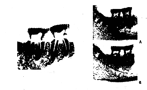
In attempting to decide whether the jaw and teeth are those of an orang or a chimpanzee, one must remember that the artificial abrasion has removed or otherwise damaged the finer details of dental morphology which distinguish these two genera. Even so, it is possible to say that they are almost certainly those of an orang. Thus, the hypsodont character of the molars and the size and shape of their pulp cavities are quite similar to those of orang teeth (Text-fig. 4), but differ markedly from those of chimpanzees which we have examined. Again, the pattern of the [241] dentine exposure corresponds very closely that that produced in our experimental abrasion of orang molars, and the numerous small fissures and pits in the central part of the occlusal surface are clearly the residual traces of crenulations of some complexity. Indeed, in one orang jaw in which we abraded the first and second molar teeth, they were found to duplicate the appearance of the Piltdown molars to a remarkable degree, not only in the extent and the contour patterns of the dentine exposures on the several cusps, in the size, depth and abrupt margins of the central basin, and in showing an approximately similar residuum of crenulations, but also in the general proportions of the crowns as a whole (including their height as measured above the enamel margin). See Pl. 27.
Text-Fig. 4. Radiographs of (A) the Piltdown molar teeth, compared with (B) those of the female orang mandible shown in Pl. 27, fig. 2. The orang is evidently more fully mature than the fossil specimen, as shown by the fact that the mandible as a whole is a little more robust. (For a radiograph of orang molars showing pulp cavities of practically identical size and shape with those of the Piltdown teeth, see Weidenreich, 1937, fig. 320). [X-rays by P. E. Purves (A) and G. M. Ardran (B).]
(II) The "Turbinal Bone"
In the supplementary note on the discoveries at Piltdown (Dawson & Woodward, 1914), Dawson stated "I saw two human nasal bones lying together with the remains of a turbinated bone beneath them in situ. The turbinal, however, was in such bad [242] condition that it fell apart on being touched, and had to be recovered in fragments by sieve ; but it has been pieced together satisfactorily . The only other reference to this find was made by Woodward in the same communication, and is limited to the following statement: "The remains of a turbinal found beneath the nasal bones are too much crushed and too fragmentary for description ; but it may be noted that the spongy bone is unusually thick, and has split longitudinally into a series of long and narrow strips." No reason was given for identifying the fragments as those of a turbinal (it is clear that a maxillo-turbinal bone is meant to be indicated by this term), except presumably by implication its proximity "in situ " to the nasal bones. Moreover, contrary to Dawson's statement, the tiny fragments (eight in number) are separate and it is not possible to fit them together to form a. complete bony element.
From an examination of these fragments it is clear that, whatever their true identification may be, they are certainly not those of a turbinal bone. They show none of the characteristic features of the maxillo-turbinal (such as its extreme thinness or its pitted and cellular texture). On the contrary, the fragments are relatively thick and they show a longitudinally grained texture which indicates that they are composed of Haversian systems arranged in parallel formation. Presumably, therefore, they are derived from the shaft of a limb bone (probably of some small animal), but their precise identification is indeterminate.
_____________________________________________________________________________
1 The large size of the pulp cavity had been noticed many years ago by Lyne (1916), but he sought to explain the combination of this feature with the severe wear on the assumption that the tooth is a deciduous canine of unusual size.
2 The penetration of the pulp cavity was not evident in the radiographs of the canine reproduced in the original communication of Dawson & Woodward (1914), for the reason that they did not happen to have been taken in just the appropriate plane.
3 When the canine tooth was picked out of a heap of gravel in 1913, it was found accurately to resemble in shape the plaster model of the canine which had already been reconstructed to fit the Piltdown mandible. Commenting on the discovery in a postscript dated 16th September, 1913, Underwood (1913) wrote "The tooth is absolutely as modelled at the British Museum."
5. The Composition of the Piltdown Hominid Remains
By K. P. Oakley
Department of Geology, British Museum (Natural History)
(1) The Mandible and Teeth
[254] The discovery in the autumn of 1953 that the Piltdown mandible contained the same amount of nitrogen as fresh bone might be regarded as proving its modernity beyond all doubt. In actual fact, however, this would not have been conclusive evidence without the cross-check provided by fluorine analysis. Thus, an ulna of woolly rhinoceros (M.12575) found at a depth of 42 ft. in an Upper Pleistocene river deposit on the site of Lloyd's in the City of London (Warren Dawson, 1925) has a nitrogen content about the same as that of the Piltdown mandible. The reason for the remarkable preservation of the rhinoceros bone is that it was embedded in an unoxidized clay - an environment in which bone-protein decays very much more slowly than in the oxidizing environment of sand or gravel. In marked contrast, a fragment of mammoth femur (M.12946) found at a depth of 20 ft. on the same site, and of the same general age, but preserved in sand, was found to contain very little nitrogen. Fortunately the fluorine content of bone increases at about the same rate in sand (or gravel) and in clay. Thus the fluorine content of the rhinoceros bone is almost the same as that of the mammoth bone.
% N % F
Upper Pleistocene bones from Lloyd's site Rhinoceros in clay av. 3.4 1.1
Elephas in sand 0.1 1.3
Piltdown mandible "in gravel" 3.9 <0.03
Fresh mammalian bones 4.1 0.03
If the Piltdown mandible had occurred naturally in the shallow Piltdown gravel (early Upper Pleistocene) it should not have contained more nitrogen than the skull bones. Moreover, its fluorine content should have been greater than that of fresh bone no matter whether the matrix were clay or gravel.
In one account of finding the mandible Woodward (1948: II) wrote: "It had evidently been missed by the workmen because the little patch of gravel in which it occurred was covered with water at the time of year when they reached it." At an early stage in the present investigation it had to be borne in mind that conditions in the Piltdown gravel might have been exceptional and that reducing conditions in the basal bed had preserved collagen in the bone. Through the courtesy of the Director of the Rothamsted Experimental Station the chemical conditions in [255] the Piltdown deposits were recently examined by Dr. C. Bloomfield, who found that the "redox potential" in the basal bed indicated oxidizing, not reducing, conditions.
In March, 1952, Professor J. T. Randall and Dr. A. V. W. Martin undertook to examine the collagen in samples of the teeth by means of the electron-microscope. The first results were inconclusive, for in drilling the samples frictional heat had probably de-natured the collagen. Early this year a new attempt was made to determine the state of collagen in the mandible. A small piece of the bone was sawn out and submitted to Dr. Martin, together with a similar sample of the cranium and other selected controls. Electron-micrographs of the decalcified residue of the mandible sample revealed the presence of fairly well preserved collagen fibres with mandible sample revealed the presence of fairly well preserved collagen fibres with characteristic banding at intervals of 640 Angstrom units (Pl. 30, fig. II), whereas electron-micrographs of samples of the skull showed no trace of collagen fibres. A residue of the Lloyd's rhinoceros bone also proved to contain collagen fibres, but they were partly de-natured and showed only vague shadows of the original banding. The only fossils in which collagen fibres have been found previously are from frozen ground -- see Randall et al., 1952. Collagen is denatured at 70o - 100o C.
It has been suggested that collagen fibres may be preserved intact through the action of peat acids, but none was detected in a sample of human bone with undiminished nitrogen content from Holocene peat at Branston, Nottinghamshire, nor in a sample of human skin from the Tollund Bog, Denmark, (kindly supplied by Dr. Knud Thorvildsen of the National Museum Copenhagen).
Estimation of the fat content of the mandible, suggested by Heizer & Cook (1954:94) as a possible means of confirming its modernity, was considered impracticable with a sample of the limited size available for such a test.
In addition to the nitrogen content, the organic carbon, water and ash contents of the mandible have been determined and compared with those of selected controls. The results confirm that this boe is modern, and also show that such artificial treatment as it received has affected the organic faction only very slightly.
%N %C %H2O %Ash
Modern angulate limb-bone (4.0) 14.0 24.5 (53)
" " " 2nd sample (4.1) 10.3 21.2 –
Piltdown mandible (3.9) 14.5 25.0 (61)
Piltdown (left parietal) 1.4 0.1 17.8 62
Neolithic human skull, Coldrum 1.9 0.3 18.2 71
Lloyd's rhinoceros ulna 3.4 10.4 18.9 (67)
Lloyd's mammoth femur 0.1 2.6 10.8 (8-)
Note.–The figures in brackets are determinations made on separate samples and therefore
independent of other figures on the same line.
The dark mahogany colour of the mandible, matching that of the skull bones very closely, is relatively superficial. Drilling revealed that the interior tissue is buff to pale grey in colour, suggesting that the organic matter filing the pores of the bone prevented the iron-staining solution from penetrating deeply. When the mandible was being drilled with a dental burr to procure an adequate sample for the re-determination of fluorine, there was an odour of burning, and the ejection [256] consisted of minute shavings. When the skull bones were drilled in the same way there was no odour and the sample consisted of powder.
The mandible shows practically no radioactivity (see Tables X, XII), which is a further confirmation of its modernity.
The fluorine content of the canine and of the molars in the mandibular ramus was estimated in 1949 as {0.1%, but as the mandibular bone itself appeared to contain c. 0.2%, and as the probable experimental error on samples of the small size then tested was known to be about ± 0.2% (Oakley & Hoskins, 1950, 379-80), there seemed no reason to regard the canine or the mandible as more recent than the human cranium, with fluorine content estimated to vary from 0.1 to 0.4%. In 1953, new samples of the teeth and of the skull bones were submitted to the Department of the Government Chemist, where Mr.C. F. M. Fryd had devised a technique for estimating smaller amounts of fluorine than could be measured in 1949. The experimental error in the determination of fluorine obviously depends on the size of the sample and the amount of fluorine it contains. The fluorine content of the Piltdown skull bones was determined in l953 as 0.14 to 0.18%, and the limits of error ± 0.02%. Where the amount measured was exceptionally small, the fluorine content was recorded as less than 0.0x % , the true figure lying between 0.0x and zero. In 1953 all the determinations of the fluorine content of the Piltdown mandible and teeth proved to lie between 0.04% and zero. These results indicated that whereas the skull bones were probably prehistoric, the canine tooth, the mandible and the isolated molar were modern. This conclusion was reinforced by comparing the nitrogen content of the Piltdown bones and teeth (dentine) with that of modern and fossil specimens.
In order to eliminate any possibility that the nitrogen found in the dentine samples was not original but due to contamination of the samples, their organic carbon was also determined. The carbon/nitrogen ratio in the molars in the canine proved to be slightly lower than in the dentine of two modern teeth which were used for comparison, but approximately the same as in the organic matter of bone (2.296 ± 0.266, Cook & Heizer, 1952:4).
%N %C C/N
Modern beaver molar (old individual) 2.2 6.5 3.0
Modern orang-utan canine 3.9 12.8 3.2
Piltdown canine 5.1 12.1 2.3
Molars in Piltdown mandible 4.3 10.0 2.3
Isolated Piltdown molar 4.2 10.7 2.5
In 1950 Dr. David B. Scott of the National Institute of Dental Research, Bethesda (Maryland) undertook to examine collodion replicas of the surfaces of the Piltdown teeth, using the metal-shadowing technique which he has developed with Wyckoff (1946). After examining replicas of the buccal surfaces of the molars in the Piltdown mandible, Dr. Scott reported that "they are not readily recognizable as ancient teeth, since they show very little evidence of post-mortem damage". But, in contrast, replicas of the isolated molar and of the crown of the canine near the tip revealed [257] considerable damage. These findings agree with the results of the present detailed re-examination, that the molars in the mandible have been artificially abraded only on the occlusal surfaces, whereas in the canine and isolated molar the buccal surfaces also have been smoothed artificially.
The black coating on the canine is a paint made from a natural bituminous earth containing iron oxide, probably Vandyke brown (see p. 272). If bituminous matter were not out of place in a highly oxidized gravel it might have been regarded as a natural incrustation. It should also be recorded that Dr. Claringbull found a minute spherule of an iron alloy embedded in the coating on the labial surface of the crown.
The pulp cavity of the canine contains 19 loose sand grains, mostly radio-opaque. Some were extracted for examination and proved to be pellets of limonitic ironstone identical with those which occur in the sand-fraction of the Piltdown gravel. As seen in the radiograph of this tooth (Weiner, Oakley & Le Gros Clark, 1953, pl. 9, fig. 4), all the grains are 1-2 mm. in diameter. If they had been naturally washed into the cavity finer grade material would be expected to have entered with them, for 30% of the grains in the sand-fraction of the Piltdown gravel are less than 1 mm. in diameter. The cavity has been sealed by an ovoid grain of hard limonitic iron stone tightly wedged into the aperture of the truncated apex.
[257] (II) The Piltdown Skull Bones
As the fluorine and nitrogen content of the cranial bones were consistent with their being fairly ancient, it seemed at first that the hoax had been based on a genuine discovery of portions of a skull in the gravel, and that the animal remains and implements had been subsequently "planted" to suggest that it was Pliocene or Early Pleistocene in age. As the investigations proceeded the skull too became suspect. Dr. G. F. Claringbull carried out an X-ray crystallographic analysis of these bones and found that their main mineral constituent, hydroxy-apatite, had been partly replaced by gypsum. Studies of the chemical conditions in the Piltdown sub-soil and ground-water showed that such an unusual alteration could not have taken place naturally in the Piltdown gravel. Dr. M. H. Hey then demonstrated that when sub-fossil bones are artificially iron-stained by soaking them in strong iron sulphate solutions this alteration does occur. Thus it is now clear that the cranial bones had been artificially stained to match the gravel, and "planted" at the site with all the other finds. The presence of chromium in some of the bones is now more readily explicable, for a dichromate solution might have served to aid the oxidation of iron salts used in staining the bones.
Since all the "local Upper Pleistocene" fossils with comparable composition have proved to be fraudulent introductions, the low fluorine content of the skull indicates that it is more probably post-Pleistocene than Pleistocene in age.
In 1912, no organic matter could be detected in the small piece of the Piltdown I calvaria submitted for analysis to Mr. S. A. Woodhead, Public Analyst of East Sussex (Dawson & Woodward, 1913:I2I). The specific gravity of the powdered fragment was also measured (2.115): but neither of these determinations was significant so long as no comparison was made with the mandible. The first physical [258] comparison between the mandible and the calvaria fragments was recorded in a note dated 1925 by Dr. A. T. Hopwood, who found that their specific gravities measured in vacuo, were as follows:
Mandible 2.06: Piltdown I occipital 2.13; Piltdown II frontal 2.18.
The specific gravities of the Piltdown II occipital and isolated molar have been determined more recently as 2.20 and 2.18 respectively. If allowance is made for the density of the molars, the specific gravity of the bone of the mandible becomes 2.05. The difference between the specific gravity of the mandible and of the calvaria would possibly have been greater if the constituent apatite had not been so extensively replaced by gypsum, which is a lighter mineral, although additional iron oxide may have counterbalanced this effect.
The age of the Piltdown skull has been questioned on the score that it included nasal bones in close association (Marston 1950:293). Unless ankylosed before death the nasal bones have commonly separated even in recent burials. It was therefore difficult to understand their occurrence together in the upper disturbed layer of the gravel (Dawson & Woodward, 1914:84). However, there was always the possibility that the nasal bones did not belong to the Piltdown skull. These bones show partial replacement by gypsum, indicating that they were artificially iron-stained. To judge from their composition they were not obtained from the same source as the other cranial fragments. In drilling samples from the nasal bones the ejection consists of shavings (as when the Piltdown mandible was drilled). This is not a proof that a bone is modern, for the property of peeling under the shearing action of a rotating burr is a function of the extent to which the collagen ground-mass of the bone is preserved; and under exceptional conditions this has persisted intact even since Pleistocene times.
Percentages
F N C H2O Ash
Fresh bone 0.03 4.1 14.0 25 53
Piltdown nasal bone 0.21 3.9 20.9 27 59
Piltdown "turbinal" 0.28 1.7 6.1 25 58
Piltdown I calvaria (maxima ) 0.18 1.9 7.5 19 70 (min. 62)
Apart from the artificial staining the Piltdown nasal bones differ from normal fresh bone only in their fluorine content, which is in excess of that found in recently buried skeletons except in areas of endemic fluorosis (Bell & Weir, 1949:89), or possibly where the soil has been treated with fluorine-rich phosphate fertilizers.
The great thickness of the Piltdown cranial bones is remarkable (e.g. maximum thickness of the parietals 12 mm.), but not unique. Cross-sections of the bones show that the thickness is accounted for by an expansion of the diploe tissues; the inner and outer tables are relatively very thin. In all those palaeolithic skulls with very thick cranial walls that have been examined histologically, the tables are nearly as thick or even thicker than the diploe (Text-fig.5). In its structure and thickness the Piltdown skull can be matched exactly among some recent crania, for example a skull of an Ona Indian from Tierra del Fuego in the British Museum collection [259] (1938.8.10.2). However, such skulls are undoubtedly rare, and to find two in the same condition at one site would be most unlikely.
The thickening of the diploe in the cranium may be a reflection of a severe chronic anaemia. The late Professor S. G. Shattock, who examined the Piltdown skull from the viewpoint of a pathologist reported (1913:46):
Text-Fig. 5. Thin sections of parietal and frontal bones of Piltdown and other human skulls. a,
Piltdown I, parietal; b. Piltdown II, frontal; c, Ona Indian, parietal; d, Swanscombe, parietal; e.
Frontéchevade II, parietal; f, Gibraltar I, parietal?. a-e are drawn from original specimens; f based on drawing by Shattack (1913). X 3. del. D. E. Woodall.
"Certain details of the Piltdown calvaria . . . suggest the possibility of a pathological process having underlain the thickened condition. These are: (1) The extreme thinness of the tables; the diploe is closed in on either aspect with the thinnest compact lamina; such as can be matched . . . [an ancient Egyptian parietal is quoted] where the thickening . . . is incontestably the residue of a morbid process. (2) The presence of the elevated patches on the inner surface of certain of the fragments already detailed: in the modern adult skull such fine vascular furrows as there may have been during growth, on the inner aspect of the tabular bones, have been smoothly filled in. And (3) to this may be added the synostosis which has here and there taken place at the sutures although the age of the individual is approximately only 25 years."
The fragments of the so-called second Pltdown skull have also been artificially stained, for they contain chromium and show partial alteration to gypsum. They comprise a small piece of occipital bone and part of a right frontal of unusual thickness. The occipital fragment is not remarkable in thickness or any other morpho[260]logical feature, but its neat rectangular outline suggests that it has been trimmed to that shape. The frontal fragment is also rectangular as though broken deliberlately. This latter piece could belong to the first cranium, with which it agrees in its exceptional thickness and unusual structure, yet in its total composition it appears to have rather more in common with the occipital fragment grouped with it than with the occipital or other bones of the first skull.
Percentages
p.p.m
N C* CaCO2 P2O5 P/P2O5 Fe CaSO4 Cr e U3O8
Piltdown I: (x 100)
Left fronto-
parietal (av.) 1.1 6.8 3.9 18.7 0.8 6 + l.5 3
Left temporal 0.2 4.8 3.6 23.2 0.8 8 ++ 0.7 1
Right parietal 1.4 5.3 3.0 19.8 0.8 5 ++ 0.0 < 1
Occipital 0.3 6.8 4.5 20.8 0.7 6 ++ 0.2 2
Piltdown II
Right frontal 1.1 4.4 1.5 14.6 0.8 10 ++ < 0.1 < 1
Occipital 0.6 3.9 2.0 13.6 0.2 9 ++ < 0.1 0
* Carbon in organic fraction.
However, when the analyses are compared, it appears possible that the resemblances between the two fragments of the Piltdown II group are not original but are due to the bones having received the same chemical treatment. The frontoparietal bone of the Piltdown I group has been less extensively altered to gypsum than the others, and it shows the highest radioactivity, the highest chromium content and the highest carbonate content. The Piltdown II bones, to judge from their high iron, low carbonate and low phosphate content, received a more intensive treatment with an acidic iron salt than any of the Piltdown I bones; and it seems probable that their lower radioactivity, 1 lower chromium and content and lower carbon content also reflect differences in treatment.
The feature in the composition of bones which is least likely to be affected by the iron sulphate and chromate treatment is the fluorine/phosphate ratio, and in this the frontal of II agrees with the bones of skull I and not with the occipital which was placed with it.
These more detailed investigations therefore lend further support to the provisional conclusion reached in our 1953 report (p. 145) that the right frontal fragment originally formed part of the first skull. It is probable that the second skull, from which the thinner occipital fragment was obtained, was in a sub-fossil condition similar to that of the first skull.
The human cranial fragments (frontal, parietal and zygomata) reported to have been found by Dawson in gravel of the Piltdown terrace at Barcombe Mills appear to [261] be pieces of two or possibly three skulls. All these fragments have been artificially iron-stained, by the sulphate process, but unaided by a chromium compound. In composition they are broadly comparable with the Piltdown calvaria, but they differ from all the fragments of these, excepting the occipital of Piltdown II, in their thickness and structure, which approximate more closely to the normal.
The possibility that occipital II belonged to one of the "Barcombe Mills" skulls has been considered, but only the parietal agrees in nitrogen content, and this differs in showing a weak radioactivity which may be original since it cannot be attributed to K40 in potassium dichromate. In contrast occipital II shows no radioactivity.
_____________________________________________________________________________
1 It has been suggested that the radioactivity of some of the Piltdon bones may be correlated with traces of K40 due to their having been treated with a solution of potassium dichromate. However,
direct tests faile to demonstrate any exact correlation between the potassium content of the specimens and either their radioactivity or their chromium content. It is probable that after the bones had been stained some of the potassium ions would have been removed in solution while the chromium ions became fixed.
[271] 8. The Black Coating on the Piltdown Canine
By A. E. A. Werner and R. J. Plesters
Research Laboratory, National Gallery
Optical examination of cross-section
In a cross-section of the black coating adhering to a fragment of the root of the canine (Text-fig.6) the following layers can be distinguished:
(a) very thin scattering of a white crystalline material on the surface, thickness c. 0.015 mm.; (b) three dark brown structureless layers (the middle one almost black) probably comprising the cementum layer of the tooth, c.0.1 mm.; (c) stained outer zone of the striated dentine, pale brown semi-transparent, c.0.2 mm.; (d) very thin zone of pale yellowish staining, c. 0.015 mm.; (e) thin zone of orange pigmentation, which shows under high power a reticulate structure, c. 0.02 mm.; (f) white, semi-transparent dentine with characteristic oblique striation.
Chemical examination of cross-section
The zoned appearance of the cross-section suggested that some material from the black coating had been absorbed preferentially through different depths. The surface was treated with organic solvent but little or no solvent effect was noted. It was then treated with dilute hydrochloric acid and a drop of potassium ferrocyanide solution. Excess was washed off with distilled water. The top layers showed a dense mass of Prussian blue, but the pale brown zone below remained unchanged (except where the precipitate had floated into the cavities). Zone d was coloured a pale green. Zone e was dark blue and Prussian blue penetrated along the striations into the unstained dentine (f ).
Text-Fig. 6. Thin-section of a fragment of the root of the Piltdown canine, showing the zones of staining. x 30 (approx.)
[272] Chemical examination of scrapings of the black coating
Under the microscope the scrapings appeared as deep golden brown translucent lumps; it was difficult to distinguish between pigment and medium, but a few reddish brown pigment particles were visible. The usual resin solvents --acetone, ethyl alcohol, benzene, chloroform -- only extracted a small quantity of material from the sample. The thin cloudy ring of extracted material fluoresced faintly in ultra-violet light. After evaporation of the solvent, the sample itself was left as a hard intact mass. (Paint having an oil-resin medium often behaves in this way a little of the resin being extracted by organic solvents, yet the pain film itself remaining apparently unchanged. Bituminous surface coatings also behave in a similar manner, a little transparent material being extracted by such solvents). Aqueous ammonia had a slight solvent effect, and alcoholic ammonia slightly grater. Morpholine and alcoholic sodium hydroxide softened and gradually disintegrated the sample. The golden brown transparent material remained dissolved completely in concentrated hydrochloric aid giving a yellow solution. This solution gave a copious precipitate of Prussian blue with potassium ferrocyanide, and a strong red coloration with ammonium thiocyanate. It must therefore contain ferric iron. From the appearance and solubility of the material this would seem to indicate the presence of a transparent iron-oxide pigment.
Further scrapings were heated in a small combustion tube. Heavy brownish fumes with a tarry smell were evolved, and condensed in the form of brown droplets. This suggested the presence of some bituminous material and would also be consistent with the behaviour of the scrapings to solvents. The incombustible residue dissolved in concentrated hydrochloric acid, and gave very strong positive reaction for iron (Fe+++).
Conclusions
The black coating contains a considerable amount of a transparent brown iron-oxide pigment; it contains some organic matter which seems to be of a bituminous nature; and the solubility tests do not exclude the presence of a little oil and/or resin. It therefore seems that the black coating is a paint consisting of a natural bituminous pigment, such as Cassel Earth or Cologne Earth (Vandyke brown), which contains a fairly high proportion of iron oxide, rather than a pure iron oxide pigment mixed with bitumen. The crackle pattern of the paint and its tough, non-brittle character (as examined on the tooth itself) are consistent with the above findings, and would suggest moreover that the surface coating is not a very great age.
[276] 10. The Radioactivity of the Piltdown Fossils 1
By S. H. U. Bowie and C. F. Davidson
Atomic Energy Division, Geological Survey of Great Britain
During recent investigations into the geochemical distribution of the radioactive elements, it was demonstrated by the Atomic Energy Division of the Geological Survey that fossil bones, teeth and other phosphatic materials tend to accrete uranium by adsorption from percolating groundwaters (Davidson & Atkin, 1953). There is some evidence that the radioactivity of a fossil bone is dependent upon the geological age of the fossil, upon the permeability of the formation in which it is found, and upon the uranium content of the pecolating waters throughout the ages. In favourable circumstances the determination of radioactivity should therefore provide a means of distinguishing older, derived fossils from contemporary bones when vertebrate remains of more than one age are found in the same geoogical envionment. No intensive research on this topic has yet been undertaken since such academic studies are rather far removed from the Atomic Energy Division 's primary function of finding workable uranium deposits.
The build-up of radioactivity in older bones is due to two separate and unrelated causes–-firstly, to the longer time that these fossils have had to adsorb uranium from circulating waters and, secondly, to the increase in radioactivity as this uranium approaches secular equilibrium with its daughter elements. The adsorped uranium has at first only about one-third of the beta radioactivity of uranium in equilibrium with its daughter elements, and the content of the latter disintegration products does not reach a maximum until equilibrium is reached– i.e. until the amount of each transitory daughter element newly generated in a given time is equal to the amount lost by decay. If uranium were present in the bone in a known amount when it was buried, and were not added to or leached away throughout its later history, the radioactivity would grdually increase for a period of several hundred thousand years, and the absolute age could be deduced therefrom. Since, however, the uranium slowly accretes after burial the radioactivity measurements can be no more than a relative indication of age between different bones found in the same geological environment.
When The Solution of the Piltdown Problem was published we suggested to the Keeper of Geology that radioactivity measureents might yield additional information on the relative ages of the fossils. He thereupon made a large number of bones and teeth available for radiometric assays, the results of which aer reported in this note.
[277] The study of phosphatic materials by autoradiographic techniques (Bowie, 1951) has shown that in radioactive bones, teeth and apatite crystals the radioactivity may be concentrated towards the surface of the specimen if the latter is relatively impermeable, but if the material is porous the adsorbed uranium tends to become evenly distributed throughout. Both for this reason, and because of the high sensitivity intrinsic to the method, the radiometric analyses of the Piltdown specimens have been conducted by measurement of the beta raiation emitted from the surface of the bone, this radiation emanating from a layer about 5 mm. in thickness. The technique employs a sensitive beta counter, with a thin mica end-window placed at a fixed distance (I cm.) from the fossil under study, the assemblage being contained in a lead chamber to reduce extraneous radiation to a minmum, and connected to a scaling unit. One great advantage of the method is that it does not consume any of the fossil material, which is preserved unchanged. A disadantae, with the equipment at present available to us, is that the specimens must be small enough (less than six inches in length) to permit their introduction into the lead chamber, and large enough (about a half-inch minimum diameter) to cover the sensitive area of the end-window counter.
Because of the variation in background count due mainly to fluctuations in cosmic radiation, the auracy of the measuements (whih is a function of the total counts recorded) depends upon the length of time allowed for the determinations. The raioactivity of a relatively uranium-rich bone can be determined with reasonable precision in a few minutes, but specimens in which the radioatiivity is very feeble demand an investigation lasting one or two days. In the table below a "standard error" is given, this being a stitistical expression of the standard deviation of the background count relative to the count given by the specimen plus background. It will be observed that in certain intance the standard error exceeds the count rate, the relevant specimens being devoid of any radioactivity determinable under these experimental conditions.
From the net count per minute given by each speciment, by extrapolation from analysed standards, the radioactitity has been expressed as equvialent urania (e.U3O8). This is a measurement of the true (chemical) uranium content only if the radioactive eleents ar in secular equilibrium and if no radioactive elements other than those of the uranium series are present. In all such phosphatic materials hitherto analysed chemically, no elements of the thorium series have been found in significant amounts; but in the Piltdown specimens which have reputedly been treated with potassium chromate, part or all of the radioactivity may be due to the potassium isotope K40 if the potash salts have not been thoroughly washed out from the bone structure. One per cent K2O has a beta radioativity equivalent to about 0.0007% U2O8.
Since we have no personal knowledge of the history or provenance of the Piltdown and other speimens submitted to us, it is not appropriate that we should attempt to interpret the results of these tests. There is a strong suggestion that the radioactivity of the bones varies sympathetically with the fluorine content, particulars of which have already been published by Oakley; but since in relatively young fossil bones the adsorbed uranium cannot have reached secular equilibrium for such [278] materials radioactive measurements may be less satisfactory than fluorine determinations as pointers to age. Possibly , however, radiometric assays may be of greater value than fluorine determinations for older fossils. It should be noted that the dentine and cementum of teeth is nearly always more radioactive than the enamel (Pl. 30,, fig. 13).
The analyses which we have obtained on a group of late Tertiary and Quaternary bones, listed below, suggest a rough correlation of radioactivity with age. Although there are too many variables governing the adsorption of uranium into such materials for radioactivity measurements in themselves to form a reliable means of dating the fossils, radiometric deterinations seem likely to provide the palaeontologist with information which, considered in conjunction with other evidence, may be an important help in discerning the relative age of two or more groups of vertebrate remains found in the same geological environment. . . .
____________________________________________________________________________
1 Communicated by permission of the Director, Geological Survey and Museum.